Do you know how your vision works?
Read on to find out about the incredible properties of the eye–and how its features affect your vision.
Eyes & Vision Science Lesson
Eye Anatomy
The human eye is one of the the most complex and sophisticated sensory organs in the body.
Its unique automatic focusing system outstrips that of any camera, and its light sensitivity is ten million times greater than the best film designed so far! Before taking a look at how the eye works, let’s start with a basic overview of how it is built.
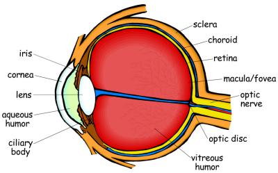
The outside layer of the eye is made up of the sclera and the cornea.
The sclera is the firm white tissue that covers all of the eye except the very front. It helps maintain the shape of the eye and protects the inner parts.
The cornea is the transparent portion at the center front part of the eye that allows light through.
A thin outer mucous membrane called the conjunctiva covers the inside of the eyelids, the cornea, and the front portion of the sclera. It helps lubricate the eye.
The middle layer of the eye contains oxygen- and nutrient-rich blood vessels, most of which are located in the layer of tissue called the choroid.
Near the front of the eye is the ciliary body, a group of muscles and ligaments that attach to the lens. These muscles change the shape of the lens as they relax and contract.
The final component of this layer is the iris, a group of muscles that controls how much light enters the eye by adjusting the opening, or pupil. The iris contains pigments that determine your eye-color.
When you look at a person’s eye, you can see parts from each of the first two layers: the “white” of the eye is the sclera, the front transparent part is the cornea, the iris is the colored part, and the pupil is the dark hole in the center.
The eye’s inner layer is composed of the retina: thin tissue that contains blood vessels and light-sensitive photoreceptor cells called rods and cones.
Every human eye contains about 120 million rods and 7 million cones.
The rods are very sensitive to low-level light, but they cannot distinguish color.
Cones need much more light to function than rods do, but they provide color information and sharp detail.
You may have noticed that in dim light color looks much less vibrant; that is because the rods that help you see in the dark are more or less “color-blind.” The retina also contains a dark pigment called melanin (also found in skin and hair cells) – this reduces reflection of light when it enters your eye.
The blood vessels and the optic nerve (the nerve that conducts electrical impulses to the brain; see the following article to learn more) connect to the retina at a spot called the optic disc.
On this disc there are no rods and cones; this is your blind spot. You don’t usually notice your blind spot, because your two eyes work together to “cover up” each other’s blind spot.
The macula is a small spot in the center of your retina. On this spot is a small pit called the fovea. When light is focused on this spot we get the sharpest image, because the fovea contains very tightly-packed photoreceptor cells. Macular degeneration is a common eye disease that is caused by the deterioration of the macula and results in partial blindness.
The three layers fill only a small part of the eye; the large middle area isn’t empty, though! The area between the cornea and the lens is filled with a transparent liquid material called the aqueous humor.The area between the lens and the retina contains a clear gel-like substance called the vitreous humor. Both of these humors help give shape to the eye and are part of the focusing process.
Your eye is a very delicate organ. The sclera and cornea protect the inner parts of the eye, but there are other protective parts as well.
Most obvious are your eyelids. With the eyelashes, your eyelids help keep outside particles from getting in your eye.
They also help spread tears, which keep the eye moist and wash away anything that gets past the eyelids. Tears are produced in the lacrimal glands and contain antibodies and anti-bacterial enzymes. The tears that your lacrimal glands produce regularly are drained away into the nasal cavity.
When you produce extra tears, though, they will spill out – this is called crying!
How Vision Works
In order to see, your eye must focus light on the retina, convert the light into electrical impulses, and send those impulses to your brain to be interpreted.
It is an amazing and complex process, but you do it constantly without even trying!
Focusing the light. When light bounces off an object and reaches the eye, it must be bent so that its rays arrive at the retina in focus.
Four different surfaces bend the light as it enters the eye: the cornea, the aqueous humor, the lens, and the vitreous humor.
When all four of these bend the light appropriately, you see a focused image of the object.
The eye can focus objects at different distances because the ciliary muscles push and pull to make the lens change shape. When you look at an object that is far away, the ciliary muscles relax and the lens has a flattened shape.
When you look at an object that is close by, the ciliary muscles are contracted and the lens is thickened. This is one of the features that makes the eye superior to any manmade camera.
To adjust a camera lens for the distance of an object, you must move the whole lens forward or back. If our eyes worked the same way, we would need long tubes sticking out of our eyes so the lenses could move back and forth.
Instead, our lenses just change shape to adjust for the distance of an object. This takes up much less room, and is probably more attractive!
In addition to focusing the light, your eye can control how much light gets in.
The colored part of your eye, called the iris, controls the size of the pupil, the opening that lets light through.
In dim light, the iris will cause your pupil to expand, allowing as much light as possible into your eye. In bright light, the iris causes the pupil to contract so that less light can enter.
Converting the light. What happens when the focused light reaches your retina? It triggers a complex chemical reaction in the light-sensitive rod and cone cells.
Rods contain a chemical called rhodopsin, or “visual purple,” and cones contain chemicals called color pigments.
These chemicals undergo a transformation that results in electrical impulses being sent to the brain through the optic nerve.
Interpreting in the brain. When the electrical impulses arrive in the visual cortex of the brain, the brain analyzes the color and light information from the rods and cones and interprets them as light.
The brain flips the image (the light was projected on your retina upside down) and fills in for the blind spot if necessary (read more on this in the science project below).
All this happens almost instantaneously, allowing you to read a book or enjoy a beautiful sunset. Some of the information from the retina is sent to the visual reflex system in your brain. This allows you to react quickly to visual threats.
If you see something coming toward your head, your visual reflex system processes this and causes you to duck before you have time to think about it!
Eyes & Vision Science Projects
Eye Chart Vision Test
A Snellen eye chart is used to determine how “normal” your vision is. It sets a standard for what most people should be able to see when they stand 20 feet away from the chart.
20/20 vision just means that when you stand 20 feet away from a Snellen eye chart, you see what a normal human being can see.
If you see 20/40, that means that when you stand 20 feet away from the chart, you see what a normal person sees standing 40 feet away from it. The higher the second number, the worse your vision is. 20/200 (you see at 20 feet what a normal person sees at 200) is the number for legal blindness in the United States.
20/20 vision isn’t perfect, it’s just “normal.” You can have better vision than 20/20. If you have 20/10 you see at 20 feet what most people see at 10. Some animals, like hawks, might have 20/2 vision!
You can use our Snellen eye chart* to compare vision within your family or with your friends.
(This will only give you an approximate idea of your vision. Your optometrist has much more precise tools to find out exactly how well you can see.)
Each line of the chart is labeled on the left side. The second to last line is 20/20.
Tape the eye chart to a wall, making sure it is in plenty of light. Stand twenty feet away from the chart and begin reading each line.
Have a family member or friend watch to see that you are reading each letter correctly. The last line that you are able to read will give you an approximate idea of your vision.
If you can read the very bottom line, your vision is 20/10! Now try covering one eye and just testing the other one. Is one eye better than the other?
Have all of your family members try reading the chart. Do some of you have better vision than others? If you wear glasses, what is your vision with them on and what is it without them?
*Instructions for downloading: The Snellen Eye Chart PDF is 11″ x 17″, so to print correctly you will need to set your print options to “tile.” Printer choices will vary, but you should do something similar to this. Open the PDF and choose Print. Under the page scaling options, select “tile all pages.” This should print the chart on four sheets of paper. You will need to trim the edges so the pieces match up, and then tape or glue them together.
(You can also order an already-printed 11″ x 17″ copy of our Snellen Eye Chart.)
Blind Spot Experiments
The spot where your optic nerve connects to your retina is called the optic disc. There are no photoreceptor cells on this disc, so when an image hits that part of your retina, you can’t see it.
This is your blind spot. You don’t notice this blind spot in every-day life, because your two eyes work together to cover it up.
To find it, draw a filled-in, 1/4″-sized square and a circle three or four inches apart on a piece of white paper.

Hold the paper at arm’s length and close your left eye. Focus on the square with your right eye, and slowly move the paper toward you. When the circle reaches your blind spot, it will disappear!
Try again to find the blind spot for your other eye. Close your right eye and focus on the circle with your left eye. Move the paper until the square disappears.
What happened when the circle disappeared? Did you see nothing where the circle had been?
No, when the circle disappeared, you saw a plain white background that matched the rest of the sheet of paper.
This is because your brain “filled in” for the blind spot – your eye didn’t send any information about that part of the paper, so the brain just made the “hole” match the rest.
Try the experiment again on a piece of colored paper. When the circle disappears, the brain will fill in whatever color matches the rest of the paper.
The brain doesn’t just match colored backgrounds. It can also make other changes to what you see. Try drawing two filled-in rectangles side by side with a circle in between them. A few inches to the right of this, draw a square.

Close your right eye and focus your left eye on the square. Move the paper until the circle disappears and the two separated bars become one bar.
How did that happen? The circle in between the bars fell on your blind spot. When it disappeared, the brain filled in for the missing information by connecting the two bars!

Here is one final experiment with your blind spot. In this instance the brain doesn’t match the blind spot with its immediate white background, but instead with the pattern surrounding it.
Draw a line down the center of your page. On one side draw a small square and on the other draw rows of circles. Color the center circle red and all the others blue.
Close your left eye and look at the square with your right eye. As you move the paper, the red circle should disappear and be replaced by a blue one!
Technology: Improving Eyesight
The general design of the human eye is practically flawless – but each individual eye isn’t.
If you are using contacts or glasses to read this article, you know that your eyes aren’t perfect.
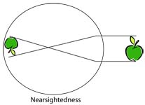
Perhaps you are nearsighted and can’t see objects that are far away very well.
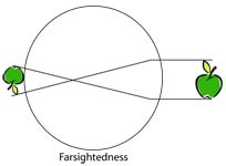
Or maybe you are farsighted and have trouble seeing things close-up. Both of these conditions occur because of the shape of the eyeball.
If your eyeball is too short, the light rays will focus the image behind your retina, instead of on it. This produces farsightedness. If your eyeball is too long, the light rays focus the image in front of the retina, making you nearsighted.
The technology of vision correction has developed over centuries.
The first known eyeglasses were made in the 13th century out of quartz set into bone, metal, or leather.
Eventually the technology for glass-blowing allowed a fine enough quality of glass to be used for lenses.
The biggest problem with these early glasses was keeping them on. It took almost 400 years before someone developed the side arms to rest on the ears!
Most people bought ready-made glasses that would have helped their vision without correcting it precisely.
For example, Benjamin Franklin had two pair of glasses, one for near and one for far. He got tired of changing them, so he cut the lenses in half and repositioned them so that he could see both near and far using the same glasses – the first bifocals!
With the advance of technology, vision-testing equipment has become more and more precise.
Now to obtain a pair of glasses, you must go to an optometrist who will determine exactly what type and strength of lenses you need.
Concave lenses are used for nearsightedness because they bend light away from the center – this stops the light from focusing too far in front of the retina.
Convex lenses are used for farsightedness because they bend light toward the center, causing the light to focus sooner so the image is not focused behind your retina.
Lenses can also be made that will correct other problems in the eye, such as astigmatism, which is an irregular curvature of the cornea.
Contact lenses are a popular alternative to eyeglasses. These lenses fit directly on the cornea, where they “float” on a layer of tears.
They were under experimentation as early as the mid-19th century, though quality and comfort left much to be desired. Now millions of people in the United States use either soft or hard lenses.
Soft contact lenses are made of flexible, water-absorbing plastics. They are more comfortable to wear than hard lenses, which are made of more rigid plastic that does not form to the eye as well. Hard lenses, on the other hand, produce a sharper image.
Some people want a more permanent solution to their vision problems. In recent years, procedures such as LASIK (laser-assisted in-situ keratomileusis) surgery have been developed to remove the need for external lenses like glasses and contacts.
While external lenses change how the light is bent so that it focuses on your retina, laser surgery reshapes the cornea itself.
The process involves a tightly focused beam of ultraviolet light, called an excimer laser. The surgeon first uses a sharp scalpel to cut a flap in the top layer of the cornea, then directs the laser into the middle layer.
As the laser pulses onto this surface, it vaporizes a microscopic portion of the cornea. By controlling the number and location of the pulses, the surgeon controls how much of the cornea is removed.
Noteworthy Scientist: Charles Bell (1774-1842)
Do you ever wonder how great artists can paint a human face that looks perfectly realistic? One of Charles Bell’s contributions to art was an anatomy textbook especially for artists, called Essays on the Anatomy of Expression in Painting.
Charles Bell was an artist himself, as well as a surgeon and anatomist. He was born in Edinburgh, Scotland, the son of a Church of England minister. His older brother John was a surgeon, author, and teacher of anatomy at the University of Edinburgh.
Studying with his brother, Bell developed both his artistic talent and his medical knowledge. After he graduated from the University with a degree in medicine, Bell assisted in teaching his brother’s anatomy class and publishing a four-volume Anatomy textbook.
Eventually Bell moved to London where he did extensive research on nerves, wrote many books and treatises, opened a school of anatomy, and worked as a surgeon.
In 1815 he cared for the wounded after the bloody battle of Waterloo, his skill in surgery holding him in good stead.
His battlefield experience led him to create illustrations of gunshot wounds to be used by surgeons.
Bell’s research on the brain and nerves proved foundational for modern neurology. He determined that nerves only sent information one way: some took sensory information to the brain, and some took commands from the brain to the rest of the body. He also traced nerves from special sensory organs (such as the eye) to specific parts of the brain.
Through all his research and medical illustration, Bell recognized the hand of a Creator. In 1836 he was invited to contribute to a collection of works “On the Power, Wisdom, and Goodness of God as Manifested in the Creation.”
He agreed, and wrote a treatise called The Hand; its Mechanism and Vital Endowment, as Evincing Design.
Bell was knighted by King William IV in 1831, and in 1835 he accepted a position as professor of surgery and returned to Scotland.
He continued to work in his field up until his death in 1842.